New Patient Evaluation “The Speech”
I call this “The Speech”. In specialty care we see the same conditions over and over, so I tend to go through this pretty quickly in the office. Excluding abdominal pain and headaches which we don’t treat, the vast majority of pain is caused by muscles, joints or nerves. Muscles can be painful but are not serious and rarely a long-term source of pain or disability. People use anti-inflammatory medicines like Motrin or Aleve, muscle relaxants, heat or ice, stretching or physical therapy, or chiropractic. We don’t see a lot of this in an interventional spine clinic.
So, it’s mainly joints or nerves and both have two things that contribute. There is underlying structural degeneration which happens slowly over years and decades, and this can be seen on imaging tests of X-rays and MRI’s. What can’t be detected on any test is inflammation. Typically, when the pain starts there was something that began an episode of inflammation and then the degeneration that was already there with no symptoms along with the inflammation which is new, together causes the pain. There is no test that can detect or measure this inflammation, so we start the treatment by doing what is known to be needed to resolve the inflammation, then if there are any residual symptoms it is from the structure. People are dramatically better, usually completely better by resolving the inflammation and that is done with injections under X-ray guidance. Medications treat the symptoms of pain; we are trying to treat the source and then there is no pain.
The medicine injected is steroids, but ours is more potent than the commonly known ones like prednisone or cortisone. It is also preservative free, so it is safe to use in the spine and skeleton. When people take oral steroids, the medication is distributed throughout the whole body so only a small amount is at the source of the pain and even if it helps it rarely lasts. The trick is getting the steroids placed at the location of the problem and that is done with X-ray guided injections.

There are four ways we can determine what the source of the pain is. There is the location of pain, physical exam, test results and most importantly is the modifying factors. There is more than one condition that can cause either back and leg pain or neck and arm pain. When it is skeletal and joints pain is most severe standing and walking because that is when there is the most pressure on the joints. When it is a spinal source of back and leg pain it is usually the most severe sitting because that is when there is the highest pressure in the intervertebral discs. We use the location of pain, modifying factors and test results to determine the source of the pain and then do injections under X-ray guidance for treatment.
When joints are the source of the pain usually a single injection is sufficient to resolve the inflammation. That is because these are synovial joints and are contained within a fibrous capsule, so once the medication gets there it stays there. Because of the capsule there is an inside and an outside and this is why people can feel worse over winter. With the barometric pressure changes of storms, the capsule becomes distended and that is why people with arthritis feel worse in winter.
For neurologic pain patients are dramatically better after the initial treatment as the inflammation is reduced, but when the inflammatory episode is well established and been present over a month it takes two injections for healing. There are two types of injections for neurologic pain of spinal stenosis. One is epidural steroids. Epidurals are the same thing we do for women in labor, but rather than using numbing for delivery we use steroids for inflammation, but the procedure is the same and even easier using X-ray. Epidurals place the medicine centrally where the nerves originate, and the medicine can go both right and left and to multiple levels. The other injection is a nerve root injection where the steroids are placed where the nerves exit the spine. This can only be done if we know which nerves are the source of the pain.
Most people return after the initial treatment and the pain is no longer constant, becomes intermittent and at least half gone. With this response we repeat the same procedure. If the response is less than this, we do the other type of procedure. These are so effective we have patients with complete relief every day. However, for an individual patient we cannot predict the response because the inflammation we are treating cannot be measured on any test. So, we begin by doing what is known to resolve the inflammation, which is two shots, and if there is any residual pain then that is the portion that is from the underlying structural degeneration.

Back and Leg Pain
There are two conditions that can cause the exact same distribution of pain in the back and leg. These are the hip or the spine. When it is the hip groin pain is prominent and much more severe with walking since that is when the joint has the most pressure. The hip joint can be stressed on physical exam to determine if this reproduces the pain.
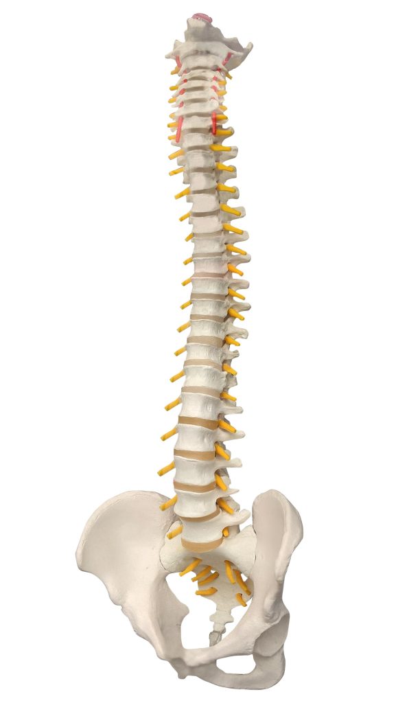
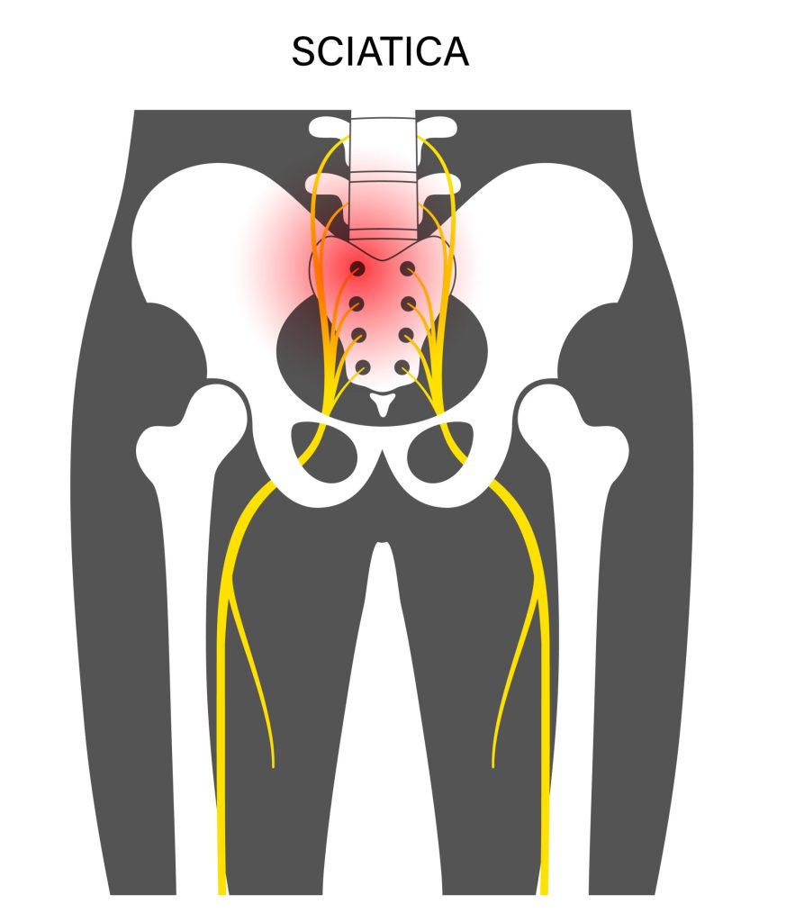
Spinal stenosis is narrowing of the spinal cord and is a very common source of back and leg pain. It is the most common condition we treat. It is often worse sitting because that increases the pressure in the intervertebral discs between the vertebra bones. Each of the nerves that exit the lumbar spine has known complete distributions. L3 is in the front of the thigh to the knee, L4 is to the outside of the calf, L5 is in the front of the shin to the big toe, and S1 is to the heel or sole of the foot. Pain in the buttock is very common with all of them and it’s not unusual to not have this full distribution. A spinal source can be tested for a physical exam and an MRI can confirm as well. This is also known as slipped disc, herniated disc, sciatica, or pinched nerve.
Whatever structural degeneration of stenosis that is seen on MRI has likely been there with little or no symptoms, and when the pain began or got worse something happened that caused the other problem of inflammation to begin. Then the narrowing that was already there and the inflammation which is new together cause the pain. The pain is dramatically better, usually completely better by getting rid of the inflammation and that is done with shots under X-ray guidance. Most patients come back after the initial treatment and the pain is no longer constant, becomes intermittent and at least half gone. The improvement is because the inflammation is less, but when the episodes of inflammation are well established and been present for over a month it takes two injections to resolve all the inflammation. With that initial response the second injection is the same as the first. If the response is less than that we do the other type of injection.
There are two types of injections for spinal stenosis. Epidural steroids are the same as epidurals for women in labor, but rather than using numbing for delivery we use steroids for inflammation, but the procedure is the same and is even easier with using X-ray. This is a common treatment if we know the pain is from a nerve, but we don’t know which nerve. With epidural steroids the medication is placed centrally where the nerves originate, and the medication can help at multiple levels and both sides.
The other type of injection is a nerve root injection, and the medication is placed where the nerves exit the spinal cord. Sometimes we know which nerve to treat based on the precise location of pain or from MRI results. There is also electrodiagnostic testing that can be obtained. When nerves are the source of pain the nerves that are causing the pain also go to certain muscles in the leg and cause cellular changes that can be detected with tiny electrodes. During the testing selected muscles are tested and when we see which muscle has these changes, then we know which nerve is causing the pain and there is a target for selective nerve root injections.
So it takes two injections, the second one may be different than the first one and there are two tests that can be obtained, an MRI or electrodiagnostics which is called an electromyelogram (EMG). These are very effective and complete relief is common, even though the structural stenosis will be ongoing. Then prevention becomes the most important education.

Neck and Arm Pain
There are two conditions that can cause the exact same distribution of pain in the neck and arm. These are the shoulder or the spine. When it is the shoulder pain is much worse with movements of the arm and range of motion is decreased. The shoulder joint can be stressed on a physical exam to determine if this reproduces the pain.
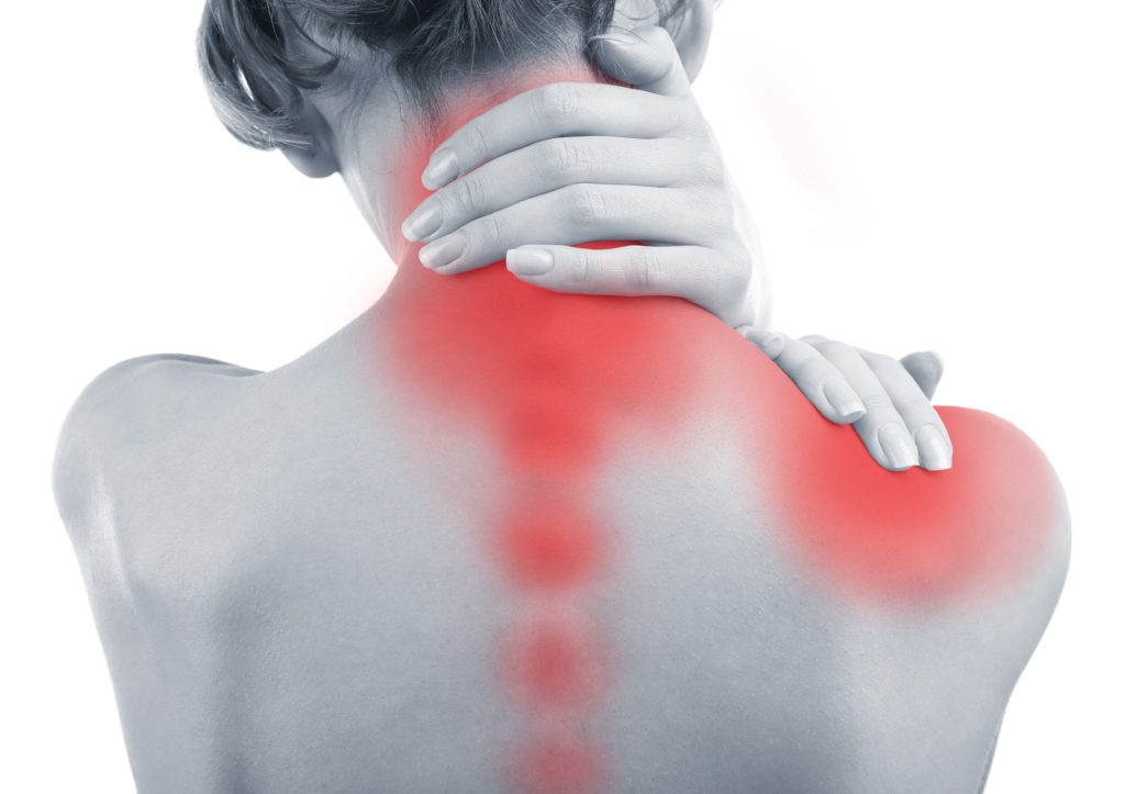
Spinal stenosis is narrowing of the spinal cord and is a very common source of neck and arm pain. It is the most common condition we treat. Each of the nerves that exit the cervical spine has known complete distributions. C4 and C5 can cause pain in the upper arm and not below the elbow, C6 to the thumb, C7 to the middle fingers, and C8 to the little finger. Pain at the shoulder blade or scapula is also common. It is not unusual to not have this full distribution. A spinal source can be tested for a physical exam and an MRI can confirm as well. This is also known as slipped disc, herniated disc, or pinched nerve.

Whatever structural degeneration of stenosis that is seen on MRI has likely been there with little or no symptoms, and when the pain began or got worse something happened that caused the other problem of inflammation to begin. Then the narrowing that was already there and the inflammation which is new together cause the pain. The pain is dramatically better, usually completely better by getting rid of the inflammation and that is done with shots under X-ray guidance. Most patients come back after the initial treatment and the pain is no longer constant, becomes intermittent and at least half gone. The improvement is because the inflammation is less, but when the episodes of inflammation are well established and been present for over a month it takes two injections to resolve all the inflammation. With that initial response the second injection is the same as the first. If the response is less than that we do the other type of injection.
There are two types of injections for spinal stenosis. Epidural steroids are the same as epidurals for women in labor, but rather than using numbing for delivery we use steroids for inflammation, but the procedure is the same and is even easier with using X-ray. This is a common treatment if we know the pain is from a nerve, but we don’t know which nerve. With epidural steroids the medication is placed centrally where the nerves originate, and the medication can help at multiple levels and both sides.
The other type of injection is a nerve root injection, and the medication is placed where the nerves exit the spinal cord. Sometimes we know which nerve to treat based on the precise location of pain or from MRI results. There is also electrodiagnostic testing that can be obtained. When nerves are the source of pain the nerves that are causing the pain also go to certain muscles in the arm and cause cellular changes that can be detected with tiny electrodes. During the testing selected muscles are tested and when we see which muscle has these changes, then we know which nerve is causing the pain and there is a target for selective nerve root injections.
So it takes two injections, the second one may be different than the first one and there are two tests that can be obtained, an MRI or electrodiagnostics which is called an electromyelogram (EMG). These are very effective and complete relief is common, even though the structural stenosis will be ongoing. Then prevention becomes the most important education.

Central Back Pain Only When Standing
Some people have pain only in their back and there is no buttock or leg pain. When there is no pain sitting and only present standing and the location is central low back there is only one condition that causes that, and we don’t even need testing. This is a result of degenerative disc disease and causes the condition of lumbar spondylosis. The degenerative disc disease can be seen on both X-rays and MRI’s. The intervertebral discs are between the vertebrae which are the bones of the spine. When they are degenerated, they no longer act as a spacer for the vertebrae and the facet joints that connect each adjacent vertebra develop spondylosis. It is essentially arthritis, but the joints are the facet joints of the spine. There is little to no pain sitting because there is no pressure on these joints, but very soon with standing there is central low back pain. These patients have difficulty standing long enough in the kitchen to make a meal, go to the mailbox, and often need to use a cart in stores. However, as soon as they sit down, they are fine.
The pain is from a combination of structural degeneration which happens slowly over years and decades, and also commonly from inflammation. Typically, when the pain starts there was something that began an episode of inflammation and then the degeneration that was already there with no symptoms along with the inflammation which is new, together causes the pain. There is no test that can detect or measure this inflammation, so we start the treatment by doing what is known to be needed to resolve the inflammation, then if there are any residual symptoms it is from the structure. People are dramatically better, usually completely better by resolving the inflammation and that is done with injections under X-ray guidance. Medications treat the symptoms of pain; we are trying to treat the source and then there is no pain.
Other treatment options if this is ineffective are prescription arthritis medications or radiofrequency ablation (RF). RF is a procedure where a probe is placed under X-ray guidance to the area where the sensory nerves are of the facet joints. These nerves are called the median branch nerves. There is a test procedure of median branch blocks where these nerves are numbed using local anesthetic and if numbing the nerve improves the pain then these patients are candidate to have the ablation. It has been called “burning the nerves”. These sensory nerve fibers are partially destroyed by the ablation so less of the information of the arthritis of the facet joints are transmitted to the brain to be interpreted as pain. This is not a permanent procedure and these nerves typically regenerate in about a year.

Posterior Hip Pain/Sacroiliac Pain
Separate from the spine is the pelvis and the joint between the sacrum bone and the iliac bone is the sacroiliac or SI joint. The location of pain is not central back, it is to the side and the back of the hip and can be the back of the thigh. Patients with this condition report difficulty rolling over in bed, sitting in a chair, offloading to one side, transitioning from sit to stand, or with stairs. There are common physical exam provocative tests that can stress the SI joint to see if this reproduces the pain.
The pain is from a combination of the structural degeneration of the joint which happens slowly over years and decades, and also commonly from inflammation. Typically, when the pain starts there was something that began an episode of inflammation and then the degeneration that was already there with no symptoms along with the inflammation which is new, together causes the pain. There is no test that can detect or measure this inflammation, so we start the treatment by doing what is known to be needed to resolve the inflammation, then if there is any residual symptoms it is from the structure. People are dramatically better, usually completely better by resolving the inflammation and that is done with injections under X-ray guidance. Medications treat the symptoms of pain, we are trying to treat the source and then there is no pain.


